選択した画像 e coli morphology under microscope 318857-What is morphology of e.coli
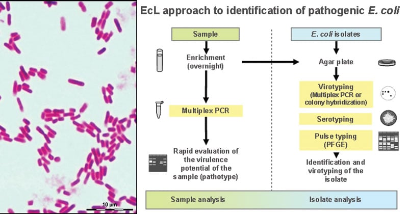
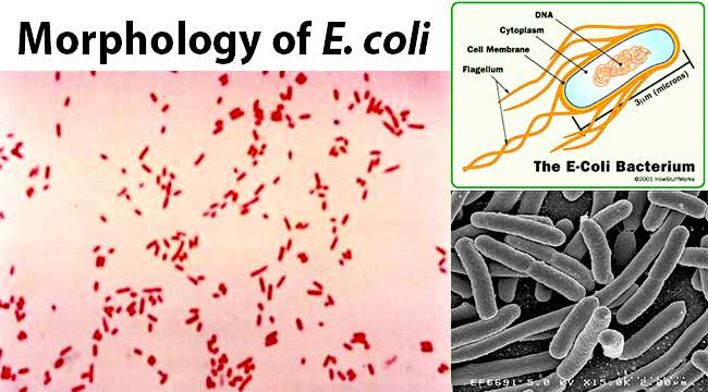
When viewed under the microscope, Gramnegative E Coli will appear pink in color The absence of this (of purple color) is indicative of Grampositive bacteria and the absence of Gramnegative E Coli Escherichia coli under 10х90х magnification using fuchsine as a dye by ElNokko (Own work) CC BYSA 40 (http//creativecommonsorg/licenses/bysa/40), via Wikimedia CommonsCULTURE REQUIREMENTS OF STAPHYLOCOCCUS AUREUSHello Viewers !!!My Name Is Kavindu Lakmal , Medical Laboratory Science Student From University Of Peradeniya I hope this video will helpful for your studi
Aph162 Report 1
What is morphology of e.coli
What is morphology of e.coli-Gram Staining reaction – Gram ve;Bacteria Under The Microscope E Coli And S Aureus Youtube Academic Oup Com Cid Article Pdf 65 4 544 Cix356 Pdf Escherichia Coli E Coli Meaning Morphology And Characteristics Escherichia Coli Uropathogenesis In Vitro Invasion Cellular Urinary Tract Infection Wikipedia


Morphological Peculiarities Of Dna Protein Complexes In Dormant Escherichia Coli Cells Subjected To Prolonged Starvation Condensation Of Dna In Dormant Cells Of Escherichia Coli Biorxiv
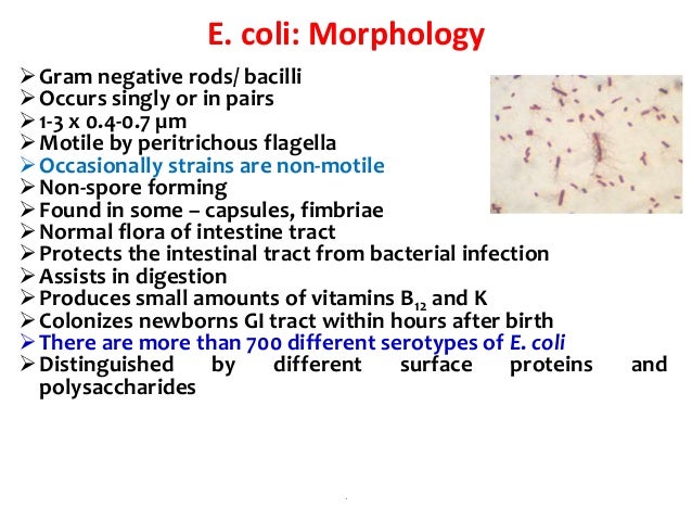
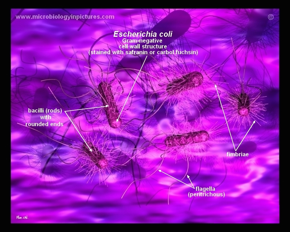
Staphylococcus aureus and Escherichia coli under microscope, morphology and microscopic appearance Bacteria under Microscope Staphylococcus epidermidis Bacteria under Microscope Gramstain Grampositive Microscopic appearance Cocci in grapelike clusters, diplococci, cocciE coli is Gramnegative straight rod, 13 µ x 0407 µ, arranged singly or in pairs (Fig 281) It is motile by peritrichous flagellae, though some strains are nonmotile Spores are not formed Capsules and fimbriae are found in some strainsBacteria Under The Microscope E Coli And S Aureus Youtube Academic Oup Com Cid Article Pdf 65 4 544 Cix356 Pdf Escherichia Coli E Coli Meaning Morphology And Characteristics Escherichia Coli Uropathogenesis In Vitro Invasion Cellular Urinary Tract Infection Wikipedia
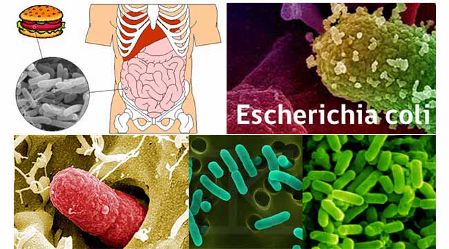
Get a second escherichia coli bacteria (e coli) stock footage at 2997fps 4K and HD video ready for any NLE immediately Choose from a wide range of similar scenes Video clip id Download footage now!Gram Staining of Micrococcus luteus,Escherichia coli, and Serratia marcescens INTRODUCTION In 14, a Danish botanist named Hans Christian Gram developed the popular staining technique known as the "gram stain"His background in studying plants at the University of Copenhagen introduced him to observing different tissues under a microscope, and thus propelled his interest in pharmacologyE coli is a gramnegative, rodshaped, facultative anaerobic bacterium that belongs to family Enterobacteriaceae It is a faecal coliform bacterium commonly found in the lower intestine of warmblooded organisms Many E coli strains are harmless, and they are a part of the normal microbiota of the gut that keeps our gut healthy However, some serotypes cause serious food poisoning, severe abdominal cramps, bloody diarrhoea, kidney failure and vomiting
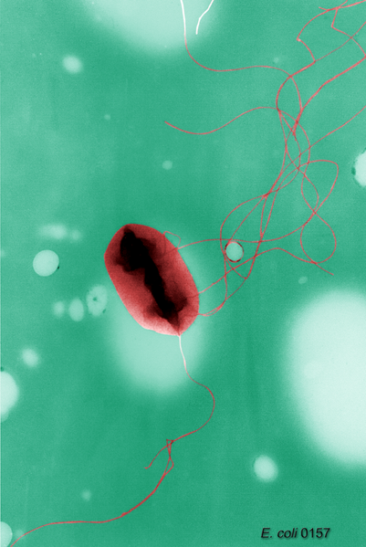
Bacteria are difficult to see under the bright field microscope Bacteria are almost colorless and therefore show little contrast with the medium in which they are suspended => To visualize bacteria various dyes are used Bacteria bear a slight negative charge => bind positively charged dyes (basic stains)MORPHOLOGY OF ESCHERICHIA COLI (E COLI) Shape – Escherichia coli is a straight, rod shape (bacillus) bacterium Size – The size of Escherichia coli is about 1–3 µm × 04–07 µm (micrometer) Arrangement Of Cells – Escherichia coli is arranged singly or in pairs Motility – Escherichia coli is a motile bacterium2 Morphology and Staining of Escherichia Coli E coli is Gramnegative straight rod, 13 µ x 0407 µ, arranged singly or in pairs (Fig 281) It is motile by peritrichous flagellae, though some strains are nonmotile Spores are not formed Capsules and fimbriae are found in some strains 3 Cultural Characteristics of Escherichia Coli


Q Tbn And9gctgrmjg8acjrbxbniknzy1qewngtoobg8cixbwsv Ok9ny02ti0 Usqp Cau
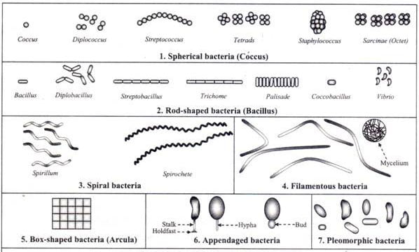


Different Size Shape And Arrangement Of Bacterial Cells
Among the most complex virions known, the T4 bacteriophage, which infects the Escherichia coli bacterium, has a tail structure that the virus uses to attach to host cells and a head structure that houses its DNA Adenovirus, a nonenveloped animal virus that causes respiratory illnesses in humans, uses glycoprotein spikes protruding from itsEscherichia coli bacteria on blood agar e coli under a microscope stock pictures, royaltyfree photos & images petri dishes with culture media for sarscov2 diagnostics, test coronavirus covid19, microbiological analysis e coli under a microscope stock pictures, royaltyfree photos & imagesZSimple Microscope consists of a single lens A hand lens is an example of a simple Microscope zCompound Microscope consists of two or more lenses in series The image formed by the first lens is further magnified by another lens Bacteria may be examined under the compound microscope, either in the living state or after fixation and staining
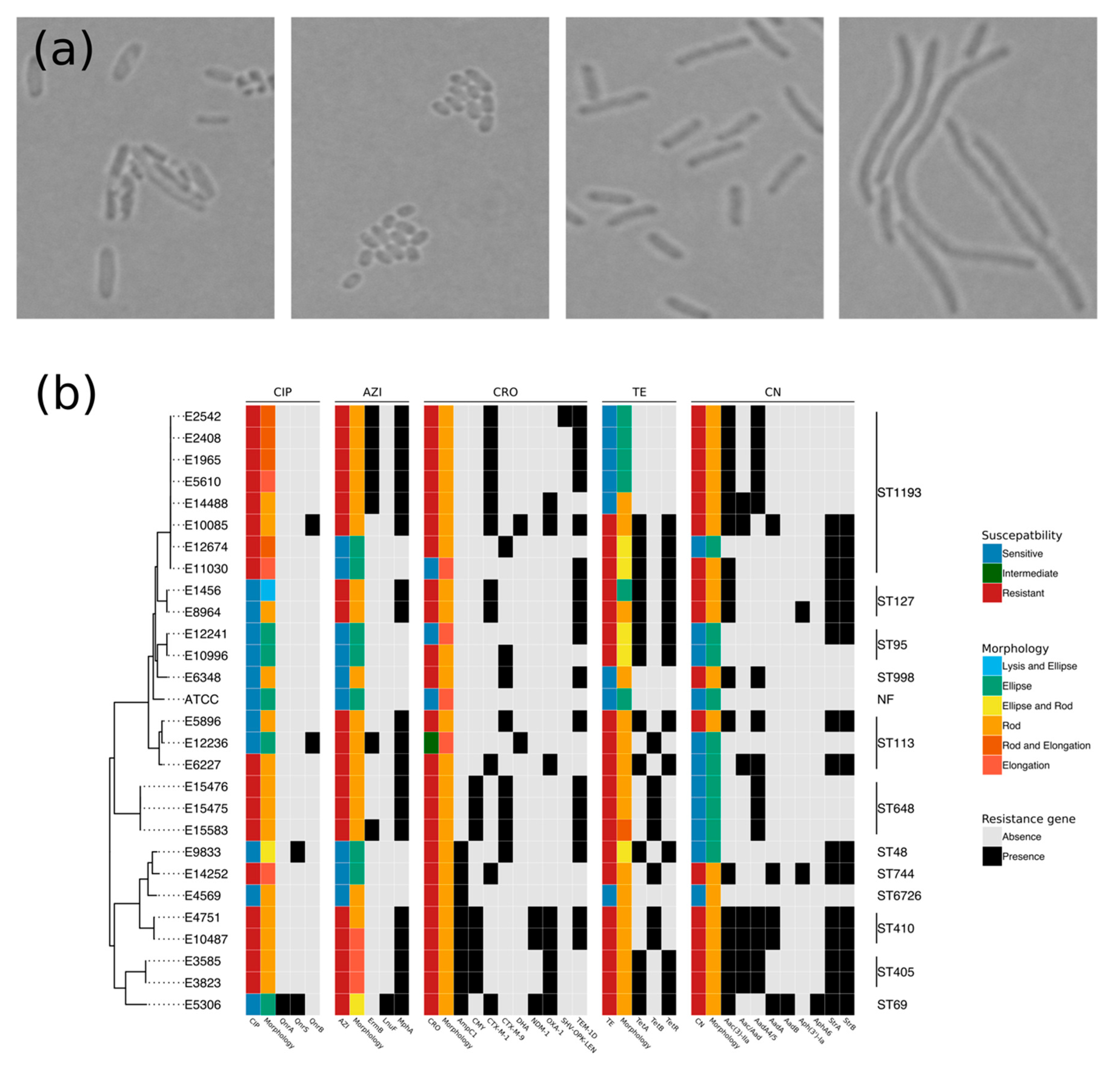


Antibiotics Free Full Text Pathogenic Escherichia Coli Possess Elevated Growth Rates Under Exposure To Sub Inhibitory Concentrations Of Azithromycin Html


Solved 1 Identify The Morphology Morphological Arrangem Chegg Com
Escherichia coli (E coli) is a species of bacteria (from the family Enterobacteraceae) that normally lives in the guts of people and animals Most E coli sCapsule – present in some strains;Escherichia coli (E coli) is a large, varied group of bacteria found in the environment, foods and lower intestines of humans and animals( Sciencing,18) When observed under the microscope the shape identified was that of a spindle or rod shape


Electron Microscopy Analysis Of E Coli And S Enteriditis Before And Download Scientific Diagram
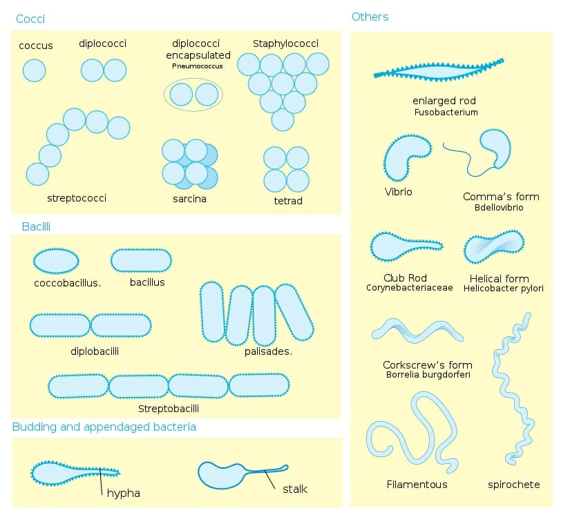


Bacteria Definition Shapes Characteristics Types Examples
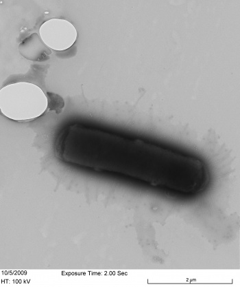
Type and morphology E coli is a Gramnegative, facultative anaerobe (that makes ATP by aerobic respiration if oxygen is present, but is capable of switching to fermentation or anaerobic respiration if oxygen is absent) and nonsporulating bacterium Cells are typically rodshaped, and are about μm long and 025–10 μm in diameter, with a cell volume of 06–07 μm 3MORPHOLOGY OF STAPHYLOCOCCUS AUREUS Shape – Round shape (cocci) Size – 1 micron (diameter) Arrangement of cells – Grapelike clusters;Examples of Bacteria Under the Microscope Escherichia coli Escherichia coli (Ecoli) is a common gramnegative bacterial species that is often one of the first ones to be observed by students Most strains of Ecoli are harmless to humans, but some are pathogens and are responsible for gastrointestinal infections They are a bacillus shaped bacteria that has a very fast growth (they can double every minutes), which is one of the main reasons they are used in research


Escherichia Coli Colony Morphology And Microscopic Appearance Basic Characteristic And Tests For Identification Of E Coli Bacteria Images Of Escherichia Coli Antibiotic Treatment Of E Coli Infections


Antigen 43 From Escherichia Coli Induces Inter And Intraspecies Cell Aggregation And Changes In Colony Morphology Of Pseudomonas Fluorescens Journal Of Bacteriology
The microscope used to determine bacterial cell morphology is A) Compound light microscope B) Dissecting microscope A) Compound light microscope Under the microscope, rods are observed randomly scattered on the field The grouping pattern is A) Cluster E) E Coli B) Corynebacterium;Small bacilli (such as Escherichia coli) that are dividing or have just divided by binary fission will look similar to cocci Look carefully for bacilli that are not dividing and are definitely rodshaped as well as bacilli in the process of dividing to confirm the true shape Bacilli do not divide so as to form clustersMost strains of E coli are rodshaped and measure about μm long and 0210 μm in diameter They typically have a cell volume of 0607 μm, most of which is filled by the cytoplasm Since it is a prokaryote, E coli don't have nuclei;
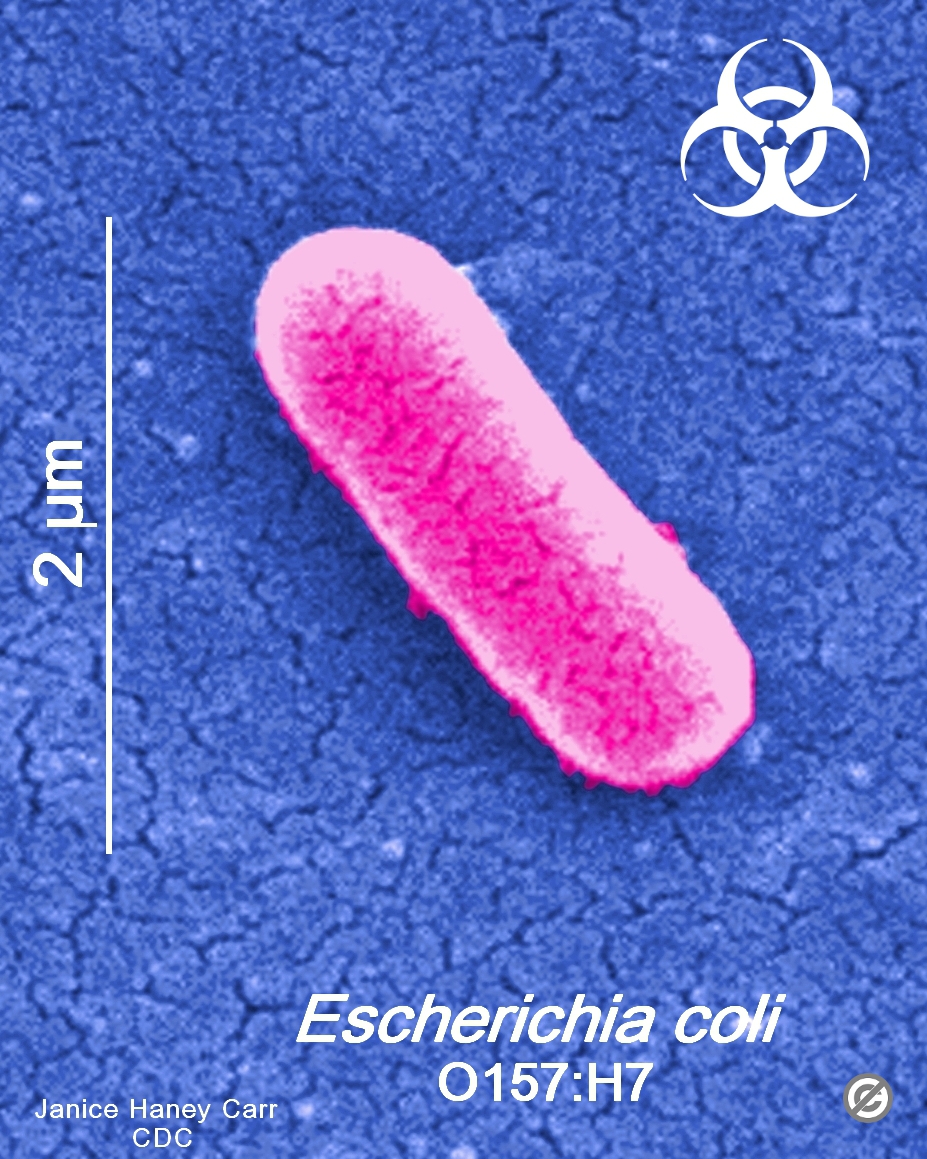


Usa Minnesota Firm Recalls Ground Beef Products Due To Possible E Coli O157 H7 Contamination Food Law Latest


Action Mode Of Bacteriocin Bm19 Against Escherichia Coli And Staphylococcus Aureus Sciencedirect
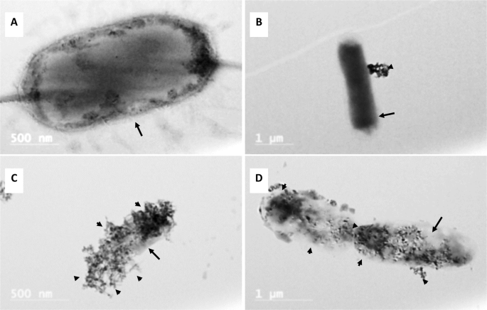
Use a dissecting/stereoscopic microscope for more detail Place the plate RIGHTSIDE UP on the stage, leaving the petri dish cover ON (Otherwise, your culture will become contaminated) There are 2 lenses on our scopes—10X and X the black lens knob is on the right side of the head of the microscopeInstead, their genetic material floats uncovered, localized to a region called the nucleoidBacteriophages, recovered from beef cattle environment and specifically targeting Escherichia coli O157H7, were examined for their physiological and morphological characteristics Degree of bacterial lysis and host range of isolated bacteriophages was determined against 55 isolates of E coli O157H7 Morphology of phages was examined under transmission electron microscope


Bacteria Under The Microscope E Coli And S Aureus Youtube


What Does An E Coli Bacteria Look Like Under A Microscope Quora
In liquid culture media like peptone water and Nutrient broth, uniform turbidity is produced which is further analyzed for the morphology (under the microscope), gram reaction, biochemical tests, and staphylococcus specific tests That's all about the Morphology & Cultural Characteristics of Staphylococcus aureusEscherichia coli, often abbreviated E coli, are rodshaped bacteria that tend to occur individually and in large clumps E coli are classified as facultative anaerobes, which means that they grow best when oxygen is present but are able to switch to nonoxygendependent chemical processes in the absence of oxygenCapsule – present in some strains;


Morphological Peculiarities Of Dna Protein Complexes In Dormant Escherichia Coli Cells Subjected To Prolonged Starvation Condensation Of Dna In Dormant Cells Of Escherichia Coli Biorxiv


Escherichia Coli Morphology Lab Diagnosis Notesmed Notesmed
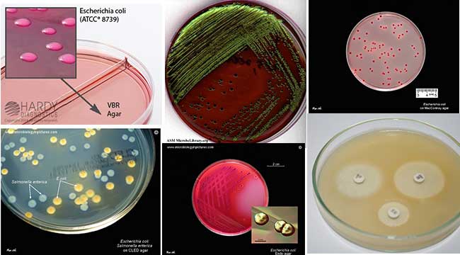
Scanning electron microscopy studies on morphology of E coli harboring pBHR68 and CRISPRi systems targeting various ftsZ gene sites, respectively E coli (pBHR68CRISPRi systems) were cultivated in Luria Bertani medium containing 2% (w/v) glucose at 37 ºC for 48 h under induction of aTc after 12 h as described in Section 2Many bacteria look like E coli when examined under the microscope (if not stained Enterobacteriaceae, Bacillus, cornyeforme bacteria, they all appear like rods, although the shape differs)This study shows how the macroscopic observation of the colonies, in particular of the colonies of Escherichia coli isolated on MacConkey agar from various clinical specimens, allowed to identify 5


Genus Escherichia Coli


Escherichia Coli E Coli An Overview Microbe Notes
Bacteria species E coli and S aureus under the microscope with different magnifications Bacteria are among the smallest, simplest and most ancient livingBacillus Cereus Morphology B cereus is classified based on how it looks under a microscope, just like most other bacteria species you normally hear about E coli contaminating differentInstead, their genetic material floats uncovered, localized to a region called the nucleoid


Gram Stain Wikipedia


How E Coli Bacteria Look Like
A pure culture of E coli would not produce two different colony types) Inability to distinguish individual cell morphology For example, all species of Staphylococcus would look the same under the microscope after simple staining)The length can range from 110 µm for filamentous or rodshaped bacteria The most wellknown bacteria E coli, their average size is ~15 µm in diameter and 26 µm in length In this figure The size comparison between our hair (~ 60 µm) and E coli (~1 µm) Notice how small the bacteria areE Coli E Coli under the microscope at 400x E Coli (Escherichia Coli) is a gramnegative, rodshaped bacterium Most E Coli strains are harmless, but some serotypes can cause food poisoning in their hosts The harmless strains are part of the normal flora of the gut Learn more about E Coli here Helicobacter Pylori


Q Tbn And9gcqkye60ou Johpr02n Mbv1fferrjpdh Lnct7ymdf5qhyia1ld Usqp Cau


Characterization And Potential Mechanisms Of Highly Antibiotic Tolerant Vbnc Escherichia Coli Induced By Low Level Chlorination Scientific Reports
Immunofluorescent and electron micrographs revealed the disrupted distribution and localisation of specific tight junction proteins (Zo1 and Claudin1) and actin filament in E coli infected Caco2 cells that ultimately resulted in deformed cellular morphology Taken together, E coli K12 under compromised in vitro milieu disrupted the intestinal barrier functions by decreasing the expression of important tight junction genes along with the altered distribution of associated proteins that• A basic light microscope has 4 objective lenses – 4X, 10X, 40X, and 100X The higher the number, the higher the power of magnification Once you have focused theCULTURE REQUIREMENTS OF STAPHYLOCOCCUS AUREUS


Escherichia Coli Wikipedia


Figure 3 From Influence Of Acetobacter Pasteurianus Sku1108 Asps Gene Expression On Escherichia Coli Morphology Semantic Scholar
Rod What is the minimum number of media that you would need to inoculate to determine the redcircled items on this flowchart?MORPHOLOGY OF STAPHYLOCOCCUS AUREUS Shape – Round shape (cocci) Size – 1 micron (diameter) Arrangement of cells – Grapelike clusters;Cells are typically rodshaped, and are about μm long and 025–10 μm in diameter, with a cell volume of 06–07 μm 3 E coli stains Gramnegative because its cell wall is composed of a thin peptidoglycan layer and an outer membrane
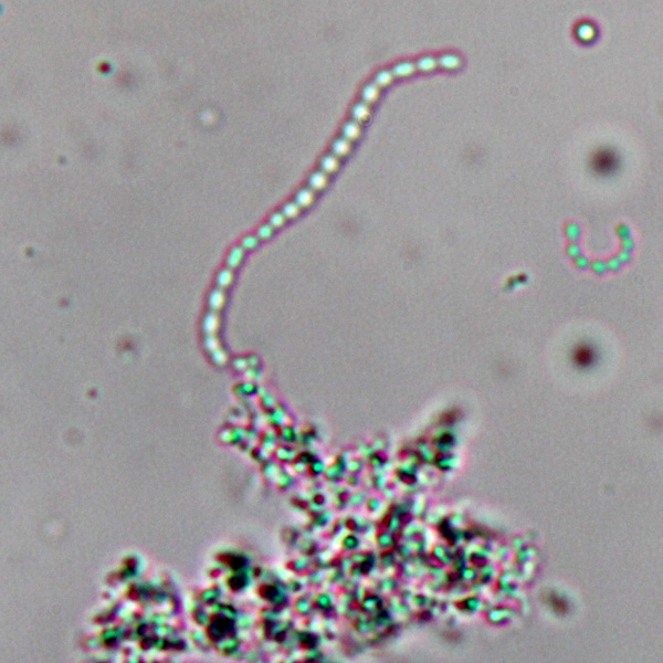


Observing Bacteria Under The Light Microscope Microbehunter Microscopy


Morphologies Of Recombinant E Coli Cells As Observed By Phase Contrast Download Scientific Diagram
It is 13 x 0407 µm in size and 06 to 07 µm in volume It is arranged singly or in pairs It is motile due to peritrichous flagella Some strains are nonmotile Some strains may be fimbriated The fimbriae are of type 1 (hemagglutinating & mannosesensitive) and are present in both motile and nonmotile strainsD) Klebsiella & E) E ColiMost strains of E coli are rodshaped and measure about μm long and 0210 μm in diameter They typically have a cell volume of 0607 μm, most of which is filled by the cytoplasm Since it is a prokaryote, E coli don't have nuclei;
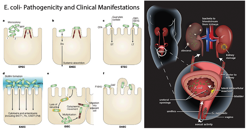


Escherichia Coli E Coli An Overview Microbe Notes


Www Myfoodresearch Com Uploads 8 4 8 5 12 Fr 19 240 Bahri 2 Pdf
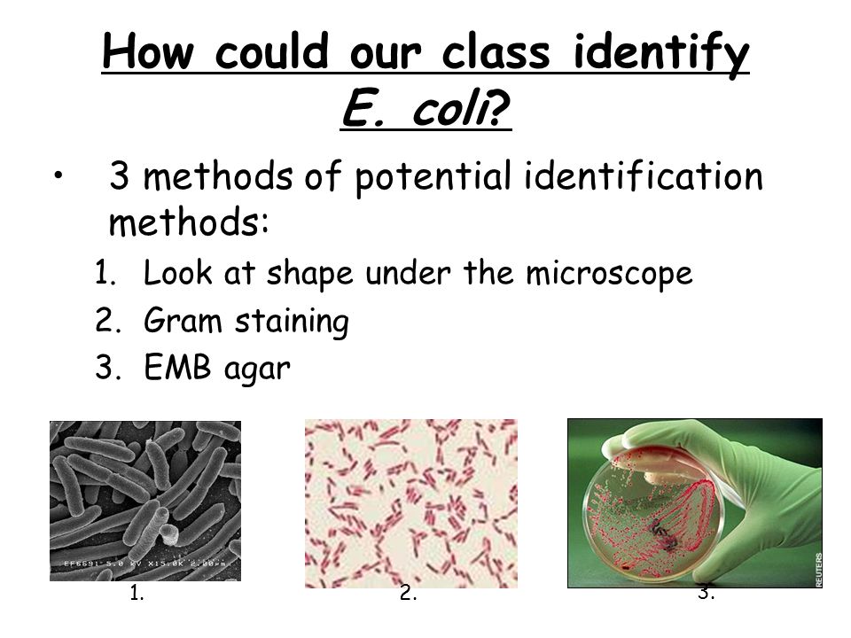
E coli is a Gramnegative rodshaped bacteria When Gram stained, the organism looks pink or red Here are a couple of pictures of a Gram stain of E coli that I did under the 100X objective lens on a standard light microscope You can of course see a lot more detail under an electron microscopeE coli would appear ___ in color and ___shaped under microscopic examination red;Entamoeba coli are a nonpathogenic ameba with world wide distribution Its life cycle is similar to that of E histolytica but it does not have an invasive stage and does not ingest red blood cells Morphology of Trophozoite The trophozoite is larger than that of E histolytica ranging from 1550μm in diameter


Aph162 Report 1


Escherichia Coli Wikipedia
Escherichia coli (E coli) is a species of bacteria (from the family Enterobacteraceae) that normally lives in the guts of people and animals Most E coli sGram Staining reaction – Gram ve;SUMMARY SUMMARY The shape of Escherichia coli is strikingly simple compared to those of higher eukaryotes In fact, the end result of E coli morphogenesis is a cylindrical tube with hemispherical caps It is argued that physical principles affect biological forms


Escherichia Coli Wikipedia


Bacteria And E Coli In Water
Microscope known bacterial characteristics of unknown Smarcescens Non motile, Cocci and Singular Gram stain ereus Gram positive Bacilli and Staphylobacillus Mluteus Gram positive, Cocci and Staphylococcus Pfluorescens Gram negative, Cocci and Triplococcus Ecoli Gram negative, rod shape and mostly exist in pairs Sepidermidis Gram positive, Cocci shape and Staphylococcus Smarcescens Gram negative, bacilli and exist in paired or chain form Table 1 shows a comparison between theA Betahemolytic colonies of Staphylococcus aureus on sheep blood agar Cultivation 24 hours, aerobic atmosphere, 37°C B Yellow colored colonies of Staphylococcus aureus on Tryptic Soy Agar Carotenoid pigment staphyloxanthin is responsible for the characteristic golden colour of S aureus colonies This pigment acts as a virulence factorStaphylococcus aureus and Ecoli under microscope microscopy of Grampositive cocci and Gramnegative bacilli, morphology and microscopic appearance of Staphylococcus aureus and Ecoli, Saureus gram stain and colony morphology on agar, clinical significance


Morphological Peculiarities Of Dna Protein Complexes In Dormant Escherichia Coli Cells Subjected To Prolonged Starvation Condensation Of Dna In Dormant Cells Of Escherichia Coli Biorxiv
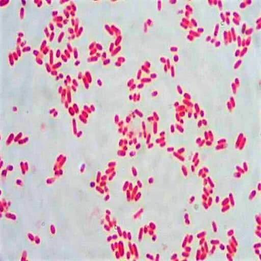


Morphology Culture Characteristics Of Escherichia Coli E Coli
E Coli Gram Stain And Cell Morphology E Coli Micrograph What Does An E Coli Bacteria Look Like Under A Microscope Quora Www Wiv Isp Be Qml Activities External Quality Rapports Atlas Bacteriology Gram Negative Aerobic And Facultative Rods Pdf
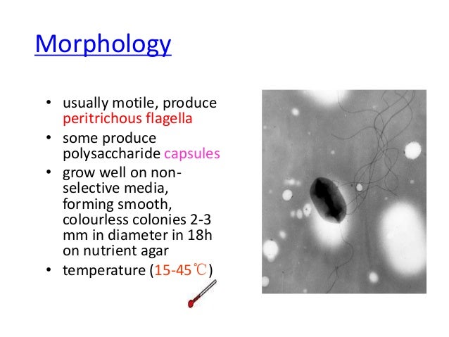


E Coli
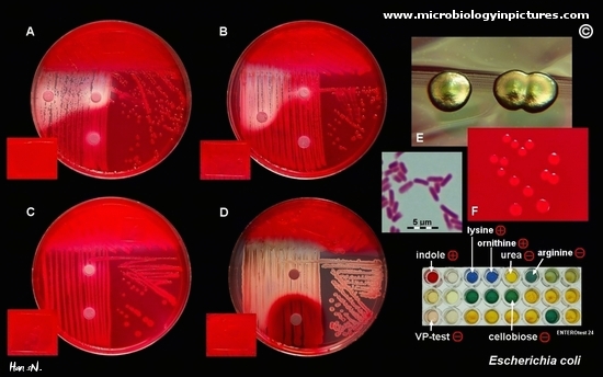


Escherichia Coli Colony Morphology And Microscopic Appearance Basic Characteristic And Tests For Identification Of E Coli Bacteria Images Of Escherichia Coli Antibiotic Treatment Of E Coli Infections


Escherichia Coli Wikipedia
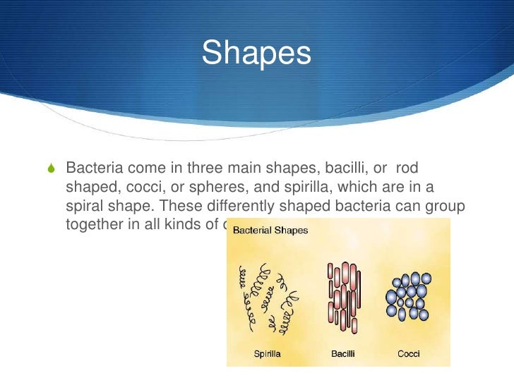


E Coli
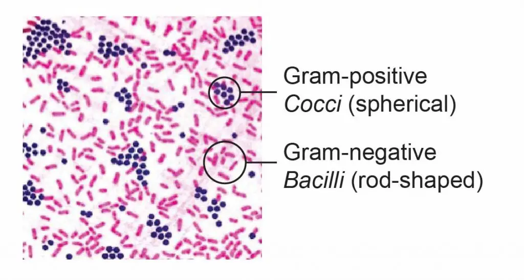


Observing Bacteria Under The Microscope Gram Stain Steps Rs Science


Introductory Chapter The Versatile Escherichia Coli Intechopen


High Precision Characterization Of Individual E Coli Cell Morphology By Scanning Flow Cytometry Konokhova 13 Cytometry Part A Wiley Online Library


Plos One The O Antigen Flippase Wzk Can Substitute For Murj In Peptidoglycan Synthesis In Helicobacter Pylori And Escherichia Coli


Lab 1


Entamoeba Coli Wikipedia


Q Tbn And9gctqlwezc G Rsexb5gmw Uv65za98k6p92dhlvblkp4mnrofyo Usqp Cau


Www Mccc Edu Hilkerd Documents Bio1lab3 Exp 4 Pdf


Escherichia Coli Responds To Environmental Changes Using Enolasic Degradosomes And Stabilized Dicf Srna To Alter Cellular Morphology Pnas


Escherichia Coli Light Microscopy


Colony Characteristics Of Escherichia Coli Is A Gramnegative Facultatively Anaerobic Rodshaped Coliform Bacterium Of The Genus Escherichia That Is Commonly Found In The Lower Intestine Stock Photo Download Image Now Istock
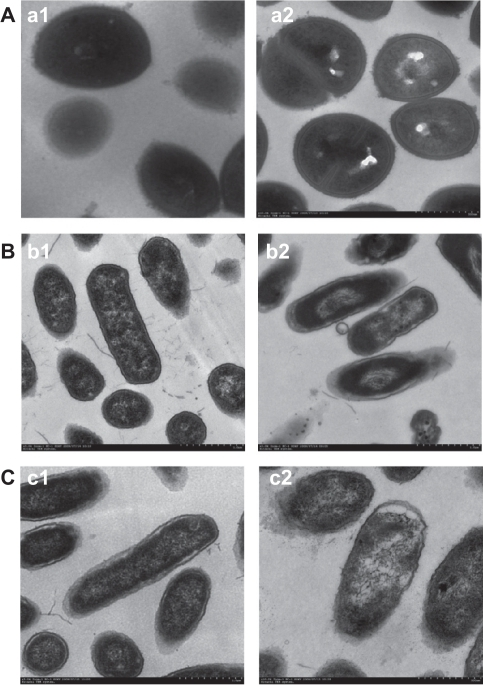


Morphology And Structure Of Bacterial Cells Under Trans Open I


E Coli


27 Earth Bacteria E Coli


Pin On Microcosmos


Colony Characteristics Of E Coli Sciencing
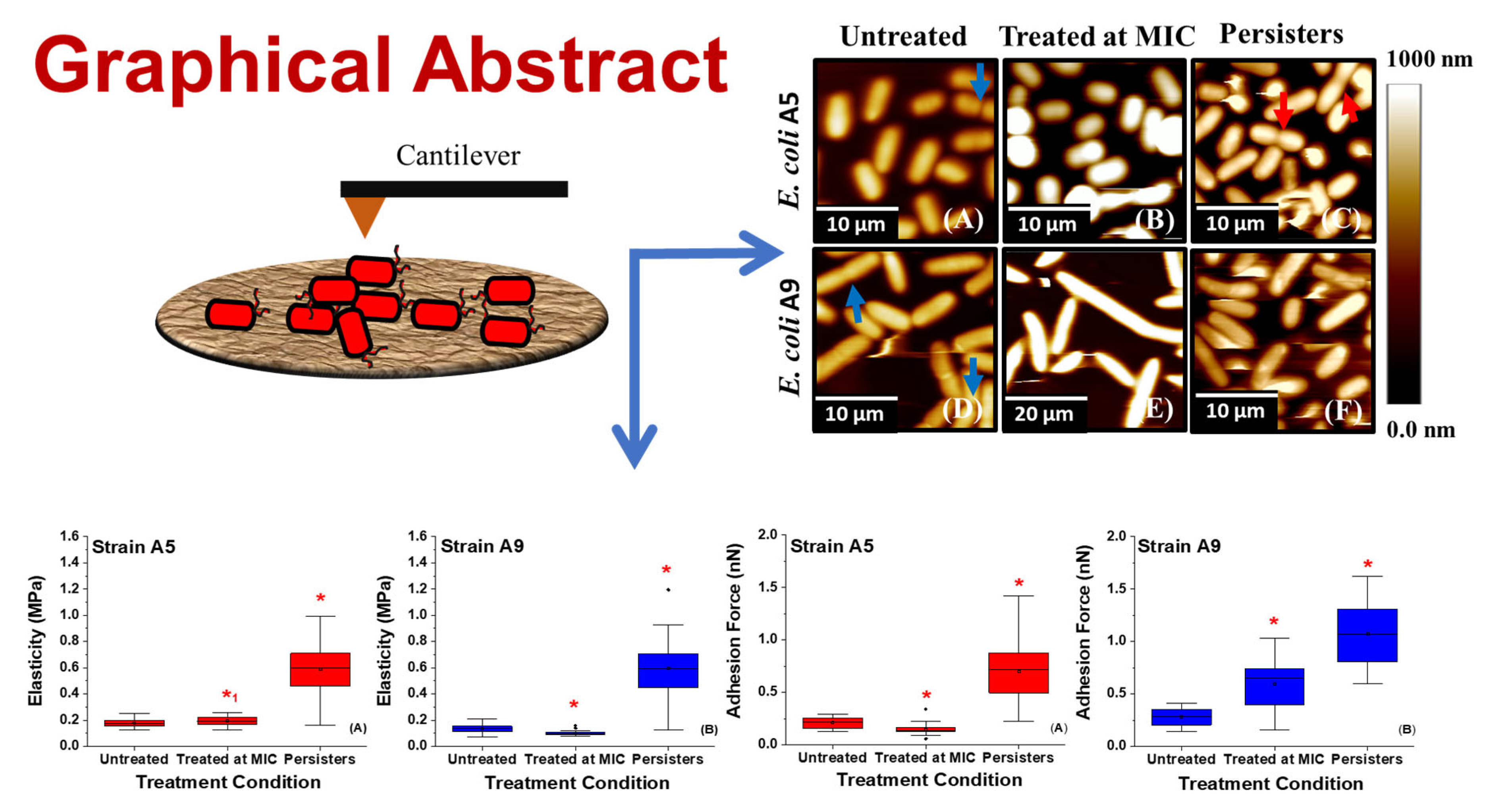


Antibiotics Free Full Text Variations In The Morphology Mechanics And Adhesion Of Persister And Resister E Coli Cells In Response To Ampicillin Afm Study


Pathogenic E Coli
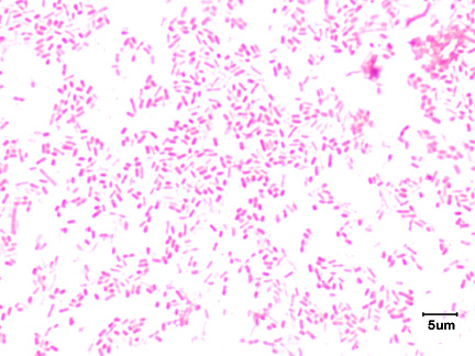


2 3b The Gram Negative Cell Wall Biology Libretexts


Morphological And Physiological Changes Induced By High Hydrostatic Pressure In Exponential And Stationary Phase Cells Of Escherichia Coli Relationship With Cell Death Applied And Environmental Microbiology


Bacteria Getting Into Shape Genetic Determinants Of E Coli Morphology Mbio
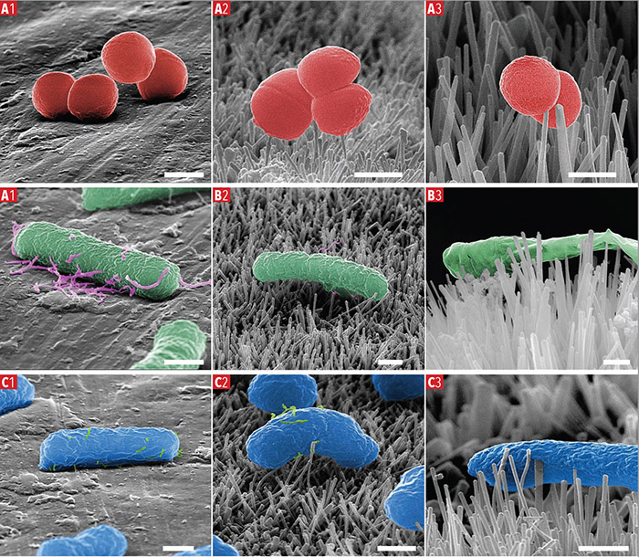


Characterisation Of Bactericidal Titanium Surfaces Using Electron Microscopy 18 Wiley Analytical Science


Escherichia Coli E Coli National Geographic Society


The Rcs Stress Response And Accessory Envelope Proteins Are Required For De Novo Generation Of Cell Shape In Escherichia Coli Journal Of Bacteriology


Intragastric Immunization Of Mice With Enterohemorrhagic Escherichia Coli O157 H7 Bacterial Ghosts Reduces Mortality And Shedding And Induces A Th2 Type Dominated Mixed Immune Response
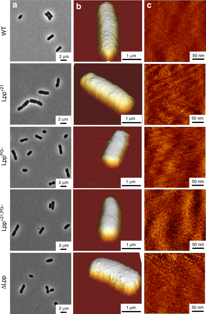


Lipoprotein Lpp Regulates The Mechanical Properties Of The E Coli Cell Envelope Nature Communications


Staphylococcus Aureus And Ecoli Under Microscope Microscopy Of Gram Positive Cocci And Gram Negative Bacilli Morphology And Microscopic Appearance Of Staphylococcus Aureus And E Coli S Aureus Gram Stain And Colony Morphology On Agar Clinical


Escherichia Coli E Coli An Overview Microbe Notes


Escherichia Coli Ecoliwiki


Staining Microscopic Specimens Microbiology


Silver Coated Magnetic Nanocomposites Induce Growth Inhibition And Protein Changes In Foodborne Bacteria Scientific Reports


Morphology Of E Coli Cells Under Microscope At 100 Magnification Download Scientific Diagram


Food Microbiology Delicious Or Ppt Video Online Download


Introductory Chapter The Versatile Escherichia Coli Intechopen
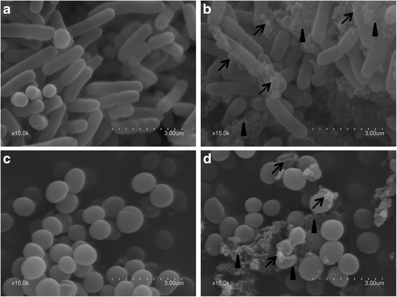


Antimicrobial Activity And Safety Evaluation Of Peptides Isolated From The Hemoglobin Of Chickens Bmc Microbiology Full Text
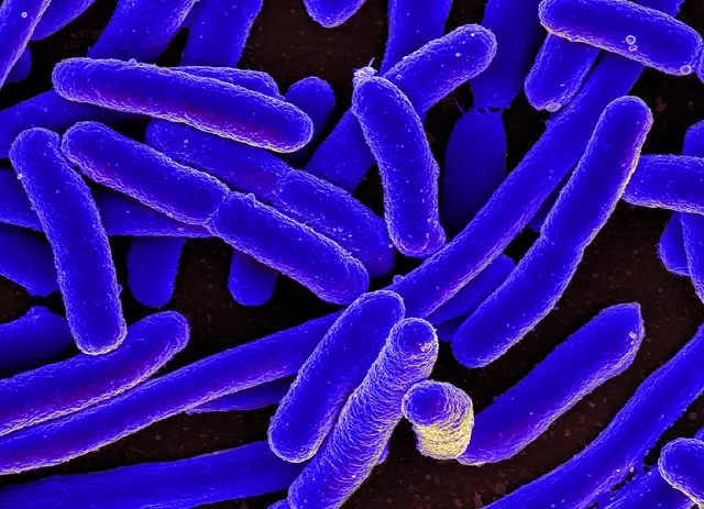


E Coli Under The Microscope Types Techniques Gram Stain Hanging Drop Method


Escherichia Coli On Macconkey Agar Colony Appearance Of Escherichia Coli Colony Morphology


Gram Stain Of E Coli Bacterium A Gram Stain Of Shows Gramnegative Download Scientific Diagram
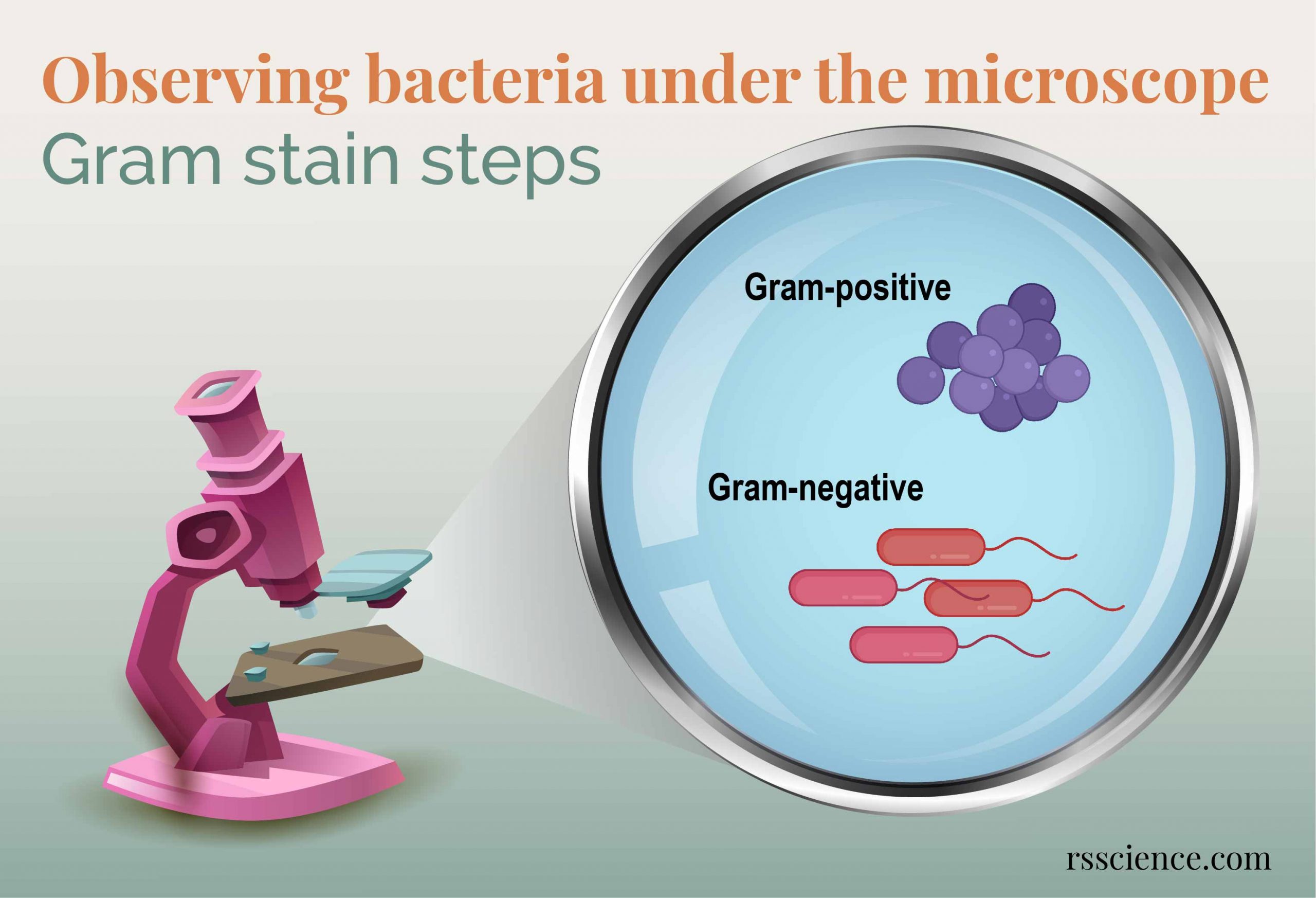


Observing Bacteria Under The Microscope Gram Stain Steps Rs Science


Growth Requirements Of E Coli And Auxotrophs Microbiology Class Video Study Com
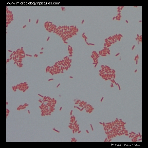


E Coli Gram Stain And Cell Morphology E Coli Micrograph Appearance Under The Microscope E Coli Cell Morphology E Coli Microscopic Picture


Enteric Bacteria List And Characteristics Medical Library


Escherichia Coli E Coli An Overview Microbe Notes


Escherichia Coli E Coli An Overview Microbe Notes


Colony Characteristics Of E Coli Sciencing


Escherichia Coli E Coli Meaning Morphology And Characteristics


Www Mccc Edu Hilkerd Documents Bio1lab3 Exp 4 Pdf


Q Tbn And9gcs3nlev0tx1pfsddwm6y9ajujk4lxjzmg7ksdxctgnipv0l50c Usqp Cau


Staphylococcus Aureus And Ecoli Under Microscope Microscopy Of Gram Positive Cocci And Gram Negative Bacilli Morphology And Microscopic Appearance Of Staphylococcus Aureus And E Coli S Aureus Gram Stain And Colony Morphology On Agar Clinical


Gram Negative Pink Colored Small Rod Shape E Coli Under Light Download Scientific Diagram


Expression Of Phi11 Gp07 Causes Filamentation In


Bacterial Characteristics Gram Staining Video Khan Academy


Figure 1 From Detection Of Multidrug Resistant And Shiga Toxin Producing Escherichia Coli Stec From Apparently Healthy Broilers In Jessore Bangladesh Semantic Scholar


Klebsiella Pneumoniae Colony Morphology And Microscopic Appearance Basic Characteristic And Tests For Identification Of Klebsiella Pneumoniae Bacteria Images Of K Pneumoniae Klebsiella Peumoniae Treatment And Antibiotics Used For K Pneumoniae


Escherichia Coli Responds To Environmental Changes Using Enolasic Degradosomes And Stabilized Dicf Srna To Alter Cellular Morphology Pnas


Effect Of The Compound No On Cell Morphology Of E Coli Cells Download Scientific Diagram


Staphylococcus Aureus And Ecoli Under Microscope Microscopy Of Gram Positive Cocci And Gram Negative Bacilli Morphology And Microscopic Appearance Of Staphylococcus Aureus And E Coli S Aureus Gram Stain And Colony Morphology On Agar Clinical


Bacteria Capsules And Slime Layers Britannica


Laboratory Perspective Of Gram Staining And Its Significance In Investigations Of Infectious Diseases Thairu Y Nasir Ia Usman Y Sub Saharan Afr J Med
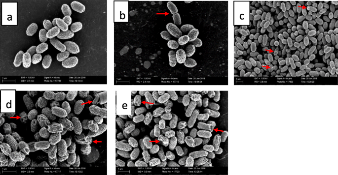


The Ultrastructural Damage Caused By Eugenia Zeyheri And Syzygium Legatii Acetone Leaf Extracts On Pathogenic Escherichia Coli Springerlink
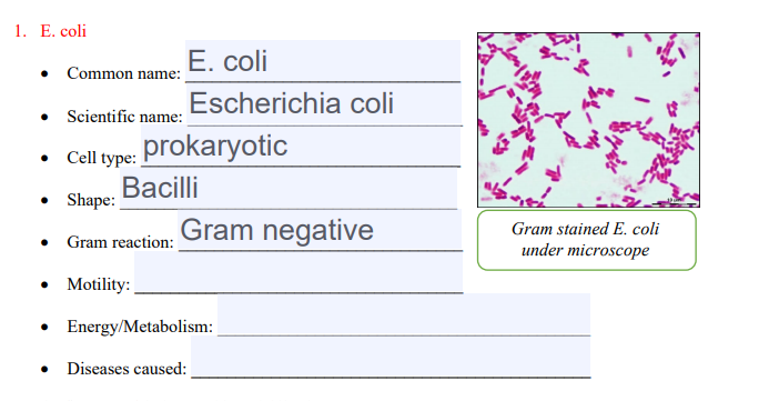


Solved 1 E Coli E Coli Common Name Escherichia Coli S Chegg Com


Scanning Electron Microscopy The Morphology Of E Coli Cells Before Download Scientific Diagram
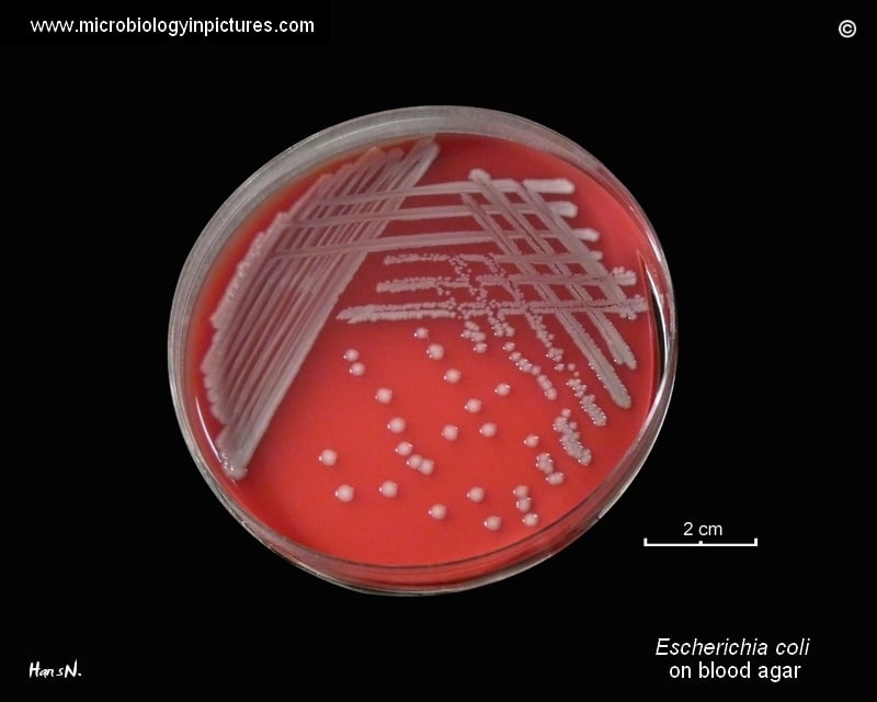


Escherichia Coli E Coli An Overview Microbe Notes


Part a K Parts Igem Org


Escherichia Coli Slide W M Science Lab Microbiology Supplies Amazon Com Industrial Scientific
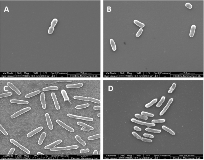


Thymol Tolerance In Escherichia Coli Induces Morphological Metabolic And Genetic Changes Bmc Microbiology Full Text


Escherichia Coli Responds To Environmental Changes Using Enolasic Degradosomes And Stabilized Dicf Srna To Alter Cellular Morphology Pnas


Staphylococcus Aureus And Ecoli Under Microscope Microscopy Of Gram Positive Cocci And Gram Negative Bacilli Morphology And Microscopic Appearance Of Staphylococcus Aureus And E Coli S Aureus Gram Stain And Colony Morphology On Agar Clinical


コメント
コメントを投稿